https://www.jst.go.jp/pr/announce/20260226/index.htmlt
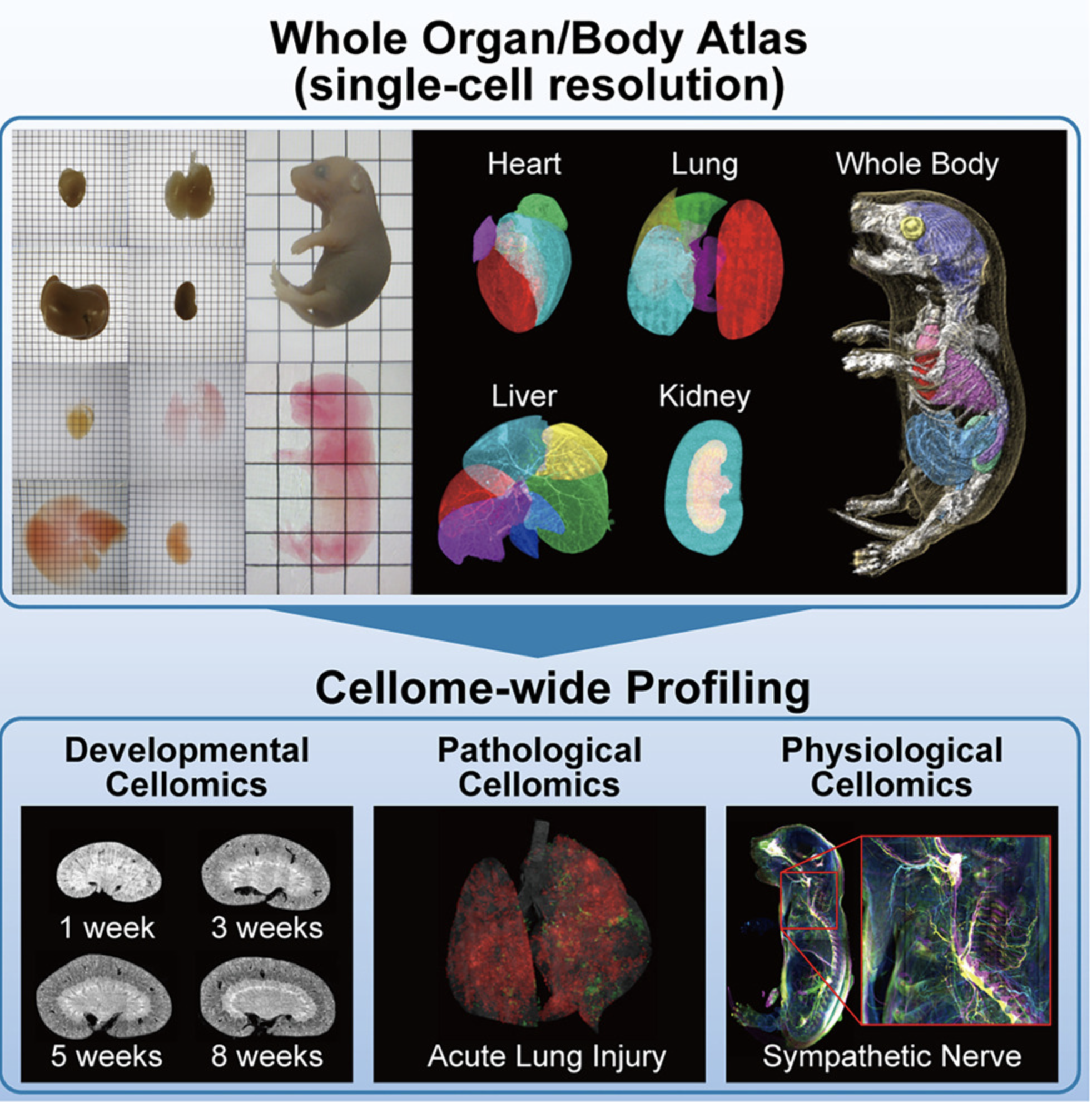
A 3D whole-cell atlas was constructed that records all cells in all organs and the entire body of mice, recording them in 3D at single-cell resolution. It serves as a foundation for quantitative analysis of developmental biology, physiology, and pathology at a whole-body scale, and is expected to be applied to next-generation 3D pathology diagnosis and drug discovery research in the future.
The research group optimized a tissue clearing method for organs and organisms (the CUBIC method) so that it can be applied to individual organs and the entire body of newborn mice, and developed a unique 3D imaging technique capable of capturing high-resolution images over a wide area. Furthermore, they extracted the positional information of each cell from the acquired 3D images and constructed a 3D atlas consisting of all cells within the entire organ or body. This makes it possible to overlay and compare cell distributions obtained from different individuals or experimental conditions based on the same reference standard.