Nikon has developed a confocal laser scanning microscope system which allows to analyze the physiological state of cells at high speed in a quantitative manner. The CRIF method (for Confocal Reflection microscopy-assisted single-cell Innate Fluorescence) acquires cell position and morphology by reflection microscopy and autofluorescence signals, which are reconstructed for each cell into a fingerprint.
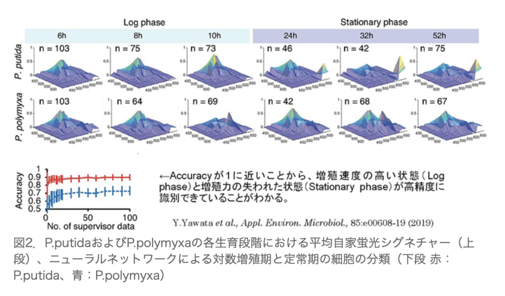
The figure below shows the change in signatures for Pseudomons putida and Polymyxa during the log and stationary phase: “Average autofluorescence signatures of P.putida and P.polymyxa at each growth stage (top row), classification of cells in logarithmic growth phase and stationary phase by neural network (bottom row) Red: P.putida, Blue: P.polymyxa”
Nikon news release, December 7, 2020
NEDO news release, December 7, 2020