https://bio.nikkeibp.co.jp/atcl/news/p1/23/02/18/10421/
A research team led by Kyoto University’s Kengo Abe and Professor Noriyuki Tsumaki (Osaka University) showed that iPS cells of cynomolgus monkeys could be differentiated into chondrocytes to prepare cartilage tissue which was successfully transplanted to other individuals. Cartilage does not contain blood vessels and is difficult for immune cells to reach, so it is low in immunogenicity, and it is expected that even cartilage derived from allogeneic iPS cells can be transplanted without immunosuppressants.
In the animal experiments, cynomolgus monkeys with cartilage defects in their knee joints were used, and allogeneic iPS cell-derived cartilage was transplanted into the defect sites. The degree of repair was evaluated at 4 and 17 weeks after transplantation. As a result, the transplanted iPS cell-derived cartilage survived to the defect site, and the degree of repair was greater the longer it was after transplantation. In addition, iPS cell-derived cartilage appears to have changed into articular cartilage after transplantation. This is because when the gene expression pattern of iPS cell-derived cartilage after transplantation was examined, marker genes for the cartilage surface layer were expressed. In this experiment on cynomolgus monkeys, it was not possible to evaluate the actual movement of joints or the improvement of subjective symptoms such as pain.
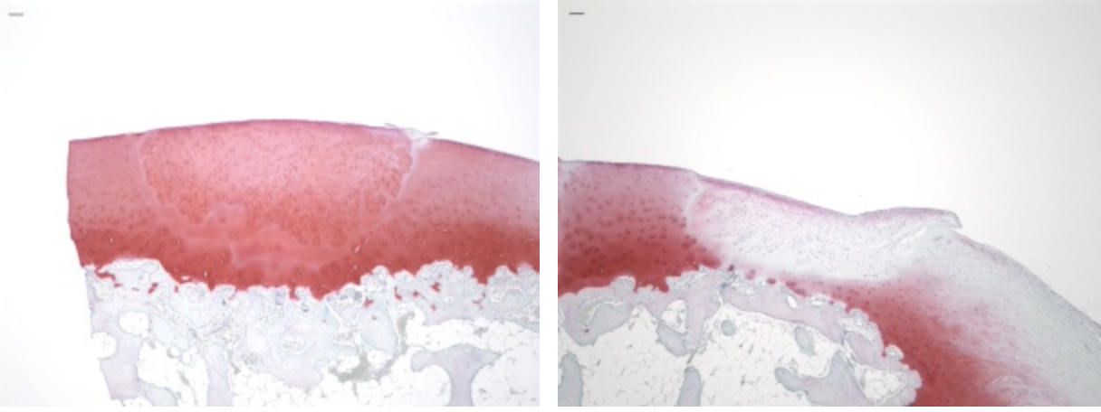
The picture show implanted iPS cell-derived cartilage (lower part) and non-transplanted (right) into the knee joint cartilage of cynomolgus monkeys with defects. The defect in the center of the image is filled with iPS cell-derived cartilage when transplanted (picture provided by Prof. Tsumaki).